
This 27 year old woman had breathlessness and chest pain since 3 years and an examination revealed only diminished breath sounds. CT scan diagnosis was “Lipomatosis”. Per op: a ‘dumbbell’ shaped fatty tumor was seen in the mediastinum that almost completely compressed both the lungs.
The huge specimen, which weighed 4.5 kg, looked like an ordinary lipoma, but on careful examination later, minute foci of grey-white areas were noted, visible to the naked eye.
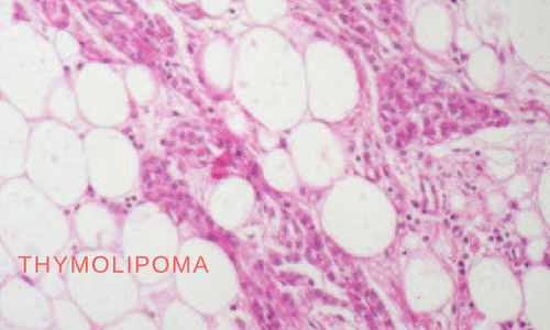
The microscopy showed islands of thymic tissue within a background of lipoma. An unusual feature of thymolipomas is the presence of cords of cuboidal epithelial cells – not found in a normal thymus (see last image).

CT scan – the thin crescent of black is the residual lung!


